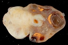
അണ്ഡാശയ ഫൈബ്രോമ
അണ്ഡാശയ ഫൈബ്രോമ, ഫൈബ്രോമ, ഒരു ബിനയിൻ സ്ട്രോമൽ ട്യൂമർ ആണ്.
| അണ്ഡാശയ ഫൈബ്രോമ | |
|---|---|
 | |
| നെവോയ്ഡ് ബേസൽ സെൽ കാർസിനോമ സിൻഡ്രോം പശ്ചാത്തലത്തിൽ കാൽസിഫൈഡ് അണ്ഡാശയ ഫൈബ്രോമയുടെ കുറഞ്ഞ മാഗ്നിഫിക്കേഷൻ മൈക്രോഗ്രാഫ്. H&E സ്റ്റെയിൻ. | |
| സ്പെഷ്യാലിറ്റി | ഗൈനക്കോളജി |

അണ്ഡാശയ ഫൈബ്രോമകൾ എല്ലാ അണ്ഡാശയ നിയോപ്ലാസങ്ങളുടെയും 4% പ്രതിനിധീകരിക്കുന്നു. [1] പെറിമെനോപോസിലും പോസ്റ്റ്മെനോപോസിലും ഇവ കൂടുതലായി സംഭവിക്കാറുണ്ട്, ശരാശരി പ്രായം ഏകദേശം 52 വയസ്സ് ആണെന്ന് റിപ്പോർട്ട് ചെയ്യപ്പെട്ടിട്ടുണ്ട്, കുട്ടികളിൽ അവ അപൂർവമാണ്. കേടുപാടുകൾ ലക്ഷണമില്ലാത്തവയാണ്. രോഗലക്ഷണങ്ങൾ ഉണ്ടെങ്കിൽ, ഏറ്റവും സാധാരണമായത് വയറുവേദനയാണ്.
ഗ്രോസ് പാത്തോളജിയിൽ, അവ ഉറച്ചതും വെളുത്തതോ തവിട്ടുനിറമോ ആണ്. സൂക്ഷ്മപരിശോധനയിൽ, കൊളാജൻ ഉത്പാദിപ്പിക്കുന്ന സ്പിൻഡിൽ സെല്ലുകളുടെ വിഭജിക്കുന്ന ബണ്ടിലുകൾ ഉണ്ട്.
കോമറ്റസ് പ്രദേശങ്ങൾ ( ഫൈബ്രോതെക്കോമ ) ഉണ്ടാകാം . അണ്ഡാശയ ഫൈബ്രോമയുടെ സാന്നിദ്ധ്യം ചില സന്ദർഭങ്ങളിൽ അണ്ഡാശയത്തെ വളച്ചൊടിക്കുന്നതിന് കാരണമാകും.
രോഗനിർണയം
തിരുത്തുകദൃഢമായ അണ്ഡാശയ മുറിവ് കാണിക്കുന്ന അൾട്രാസോണോഗ്രാഫി ഉപയോഗിച്ചാണ് സാധാരണയായി രോഗനിർണയം നടത്തുന്നത്, അല്ലെങ്കിൽ ചില അവസരങ്ങളിൽ സോളിഡ്, സിസ്റ്റിക് ഘടകങ്ങളുള്ള മിക്സഡ് ട്യൂമറുകൾ. [3] കമ്പ്യൂട്ട്ഡ് ടോമോഗ്രഫി, മാഗ്നെറ്റിക് റിസോണൻസ് ഇമേജിംഗ് എന്നിവയും ഫൈബ്രോമകൾ നിർണ്ണയിക്കാൻ ഉപയോഗിക്കാം. 16 രോഗികളുടെ ഒരു പരമ്പരയിൽ, 5 (28%) CA-125 ന്റെ ഉയർന്ന അളവ് കാണിച്ചു. ഹിസ്റ്റോപത്തോളജി സ്പിൻഡിൽ ആകൃതിയിലുള്ള ഫൈബ്രോബ്ലാസ്റ്റിക് കോശങ്ങളും സമൃദ്ധമായ കൊളാജനും കാണിക്കുന്നു. [4]
ചികിത്സ
തിരുത്തുകസാധാരണയായി മുറിവ് ശസ്ത്രക്രിയയിലൂടെ നീക്കംചെയ്യുന്നു. പ്രാഥമികമായി, ഒരു രോഗിയിൽ കണ്ടെത്തിയ മുറിവ് അർബുദമാകുമെന്ന ആശങ്കയുണ്ട്, പക്ഷേ ടോർഷൻ സാധ്യതയും രോഗലക്ഷണങ്ങൾ ഉണ്ടാകാനുള്ള സാധ്യതയും ഉണ്ട്. എന്നിരുന്നാലും, സ്ഥിരതയുള്ള ഒരു ക്ഷതം, ക്ലിനിക്കലി പിന്തുടരാവുന്നതാണ്.
അസോസിയേഷനുകൾ
തിരുത്തുകഎഡിമ ഉള്ള വകഭേദങ്ങൾ മീഗ്സ് സിൻഡ്രോമുമായി ബന്ധപ്പെടുത്താം. അവർ നെവോയിഡ് ബേസൽ സെൽ കാർസിനോമ സിൻഡ്രോമിന്റെ (ഗോർലിൻ സിൻഡ്രോം) ഭാഗമാകാം. [5]
റഫറൻസുകൾ
തിരുത്തുക- ↑ Yen, P.; Khong, K.; Lamba, R.; Corwin, M. T.; Gerscovich, E. O. (2013). "Ovarian fibromas and fibrothecomas: Sonographic correlation with computed tomography and magnetic resonance imaging: A 5-year single-institution experience". Journal of Ultrasound in Medicine. 32 (1): 13–18. doi:10.7863/jum.2013.32.1.13. PMID 23269706.
- ↑ - Vaidya, SA; Kc, S; Sharma, P; Vaidya, S (2014). "Spectrum of ovarian tumors in a referral hospital in Nepal". Journal of Pathology of Nepal. 4 (7): 539–543. doi:10.3126/jpn.v4i7.10295. ISSN 2091-0908.
- Minor adjustment for mature cystic teratomas (0.17 to 2% risk of ovarian cancer): Mandal, Shramana; Badhe, Bhawana A. (2012). "Malignant Transformation in a Mature Teratoma with Metastatic Deposits in the Omentum: A Case Report". Case Reports in Pathology. 2012: 1–3. doi:10.1155/2012/568062. ISSN 2090-6781. PMC 3469088. PMID 23082264.{{cite journal}}: CS1 maint: unflagged free DOI (link) - ↑ Yen, P.; Khong, K.; Lamba, R.; Corwin, M. T.; Gerscovich, E. O. (2013). "Ovarian fibromas and fibrothecomas: Sonographic correlation with computed tomography and magnetic resonance imaging: A 5-year single-institution experience". Journal of Ultrasound in Medicine. 32 (1): 13–18. doi:10.7863/jum.2013.32.1.13. PMID 23269706.Yen, P.; Khong, K.; Lamba, R.; Corwin, M. T.; Gerscovich, E. O. (2013). "Ovarian fibromas and fibrothecomas: Sonographic correlation with computed tomography and magnetic resonance imaging: A 5-year single-institution experience". Journal of Ultrasound in Medicine. 32 (1): 13–18. doi:10.7863/jum.2013.32.1.13. PMID 23269706.
- ↑ Parwate, Nikhil Sadanand; Patel, Shilpa M.; Arora, Ruchi; Gupta, Monisha (2015). "Ovarian Fibroma: A Clinico-pathological Study of 23 Cases with Review of Literature". The Journal of Obstetrics and Gynecology of India. 66 (6): 460–465. doi:10.1007/s13224-015-0717-6. ISSN 0971-9202. PMC 5080219. PMID 27821988.
- ↑ Tytle, T.; Rosin, D. (Sep 1984). "Bilateral calcified ovarian fibromas". South Med J. 77 (9): 1178–80. doi:10.1097/00007611-198409000-00033. PMID 6385289.