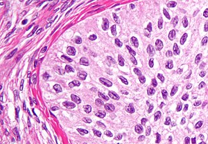
സർഫേസ് എപ്പിത്തീലിയൽ-സ്ട്രോമൽ ട്യൂമർ
ഈ ലേഖനം ഇംഗ്ലീഷ് ഭാഷയിൽ നിന്ന് കൃത്യമല്ലാത്ത/യാന്ത്രികമായ പരിഭാഷപ്പെടുത്തലാണ്. ഇത് ഒരു കമ്പ്യൂട്ടറോ അല്ലെങ്കിൽ രണ്ട് ഭാഷയിലും പ്രാവീണ്യം കുറഞ്ഞ ഒരു വിവർത്തകനോ സൃഷ്ടിച്ചതാകാം. |
അണ്ഡാശയ കോശപ്പെരുപ്പങ്ങളുടെ ഒരു വിഭാഗമാണ് സർഫേസ് എപ്പിത്തീലിയൽ-സ്ട്രോമൽ ട്യൂമർ. അവ ദോഷകരമോ മാരകമോ ആകാം. ഈ ഗ്രൂപ്പിലെ കോശപ്പെരുപ്പങ്ങൾ അണ്ഡാശയ ഉപരിതല എപ്പിത്തീലിയത്തിൽ നിന്നോ (ഭേദഗതി വരുത്തിയ പെരിറ്റോണിയം) അല്ലെങ്കിൽ എക്ടോപിക് എൻഡോമെട്രിയൽ അല്ലെങ്കിൽ ഫാലോപ്യൻ ട്യൂബ് (ട്യൂബൽ) ടിഷ്യൂവിൽ നിന്നോ ഉരുത്തിരിഞ്ഞതാണെന്ന് കരുതപ്പെടുന്നു. ഇത്തരത്തിലുള്ള മുഴകളെ അണ്ഡാശയ അഡിനോകാർസിനോമ എന്നും വിളിക്കുന്നു.[1]അണ്ഡാശയ അർബുദത്തിന്റെ എല്ലാ കേസുകളിലും 90% മുതൽ 95% വരെ മുഴകൾ ഈ ഗ്രൂപ്പാണ്. എന്നിരുന്നാലും 40 വയസ്സിന് താഴെയുള്ള ആർത്തവവിരാമം നേരിടുന്ന സ്ത്രീകളിൽ 7% കേസുകളും യുണൈറ്റഡ് സ്റ്റേറ്റ്സ് ഒഴികെയുള്ള സ്ത്രീകളിൽ മാത്രമാണ് ഇത് പ്രധാനമായും കാണപ്പെടുന്നത്.[2][3][4][5][6][7] സെറം CA-125 പലപ്പോഴും ഉയർന്നതാണ്. പക്ഷേ ഇത് 50% മാത്രം കൃത്യതയുള്ളതിനാൽ ചികിത്സയുടെ പുരോഗതി വിലയിരുത്തുന്നതിന് ഇത് ഉപയോഗപ്രദമായ ട്യൂമർ മാർക്കർ അല്ല. എപ്പിത്തീലിയൽ അണ്ഡാശയ അർബുദമുള്ള 75% സ്ത്രീകളും വിപുലമായ ഘട്ടങ്ങളിൽ കാണപ്പെടുന്നു. എന്നിരുന്നാലും, പ്രായമായ രോഗികളെ അപേക്ഷിച്ച് പ്രായം കുറഞ്ഞ രോഗികൾക്ക് മികച്ച രോഗനിർണയം ഉണ്ടാകാനുള്ള സാധ്യത കൂടുതലാണ്.[8][9][10][11] [12]
| Surface epithelial-stromal tumor | |
|---|---|
 | |
| ബ്രണ്ണർ ട്യൂമറിന്റെ ഉയർന്ന മാഗ്നിഫിക്കേഷൻ മൈക്രോഗ്രാഫ്, ഒരു തരം ഉപരിതല എപ്പിത്തീലിയൽ-സ്ട്രോമൽ ട്യൂമർ. എച്ച്&ഇ കറ.. | |
| സ്പെഷ്യാലിറ്റി | Oncology |
വർഗ്ഗീകരണം
തിരുത്തുകഎപ്പിത്തീലിയൽ-സ്ട്രോമൽ ട്യൂമറുകൾ എപ്പിത്തീലിയൽ സെൽ തരം, എപിത്തീലിയത്തിന്റെയും സ്ട്രോമയുടെയും ആപേക്ഷിക അളവ്, പാപ്പില്ലറി പ്രക്രിയകളുടെ സാന്നിധ്യം, എപ്പിത്തീലിയൽ മൂലകങ്ങളുടെ സ്ഥാനം എന്നിവയെ അടിസ്ഥാനമാക്കിയാണ് തരംതിരിച്ചിരിക്കുന്നത്. ഉപരിതല എപ്പിത്തീലിയൽ-സ്ട്രോമൽ ട്യൂമർ ദോഷകരമാണോ, ബോർഡർലൈൻ ട്യൂമർ അല്ലെങ്കിൽ മാലിഘ്നന്റ് (evidence of malignancy and stromal invasion) മൈക്രോസ്കോപ്പിക് പാത്തോളജിക്കൽ ലക്ഷണങ്ങൾ നിർണ്ണയിക്കുന്നു. ബോർഡർലൈൻ ട്യൂമറുകൾ അനിശ്ചിതത്വത്തിൽ മാരകമായ സാധ്യതയുള്ളവയാണ്.
ഈ ഗ്രൂപ്പിൽ സീറസ്, മ്യൂസിനസ്, എൻഡോമെട്രിയോയിഡ്, ക്ലിയർ സെൽ, ബ്രെന്നർ ട്യൂമർ (ട്രാൻസിഷണൽ സെൽ) എന്നിവ അടങ്ങിയിരിക്കുന്നു. എന്നിരുന്നാലും സമ്മിശ്രവും വേർതിരിക്കപ്പെടാത്തതും തരംതിരിക്കാത്തതുമായ ചില തരങ്ങളുണ്ട്.
അവലംബം
തിരുത്തുക- ↑ 1.0 1.1 Kosary CL (2007). "Chapter 16: Cancers of the Ovary" (PDF). In Baguio RN, Young JL, Keel GE, Eisner MP, Lin YD, Horner MJ (eds.). SEER Survival Monograph: Cancer Survival Among Adults: US SEER Program, 1988-2001, Patient and Tumor Characteristics. SEER Program. Vol. NIH Pub. No. 07-6215. Bethesda, MD: National Cancer Institute. pp. 133–144.
- ↑ Centers for Disease Control and Prevention, CDC WONDER. United States and Puerto Rico cancer statistics, 1999–2013 inci- dence incidence request.Available at: http://wonder.cdc.gov/ cancer-v2013.html. Retrieved December 1, 2016.
- ↑ Kashima K, Yahata T, Fujita K, Tanaka K. Outcomes of fertility-sparing surgery for women of reproductive age with FIGO stage IC epithelial ovarian cancer. Int J Gynaecol Obstet 2013;121:53–5.
- ↑ Rauh-Hain JA, Foley O, Winograd D, Andrade C, Clark RM, Vargas RJ, et al. Clinical characteristics and outcomes of pa- tients with stage I epithelial ovarian cancer compared to fallo- pian tube cancer. Am J Obstet Gynecol 2015;212:600.e1–8.
- ↑ Wright JD, Shah M, Mathew L, Burke WM, Culhane J, Gold- man N, et al. Fertility preservation in young women with epi- thelial ovarian cancer. Cancer 2009;115:4118–26.
- ↑ Melamed A., Rizzo A.E., Nitecki R., et al All-cause mortality after fertility-sparing surgery for stage i epithelial ovarian cancer. Obstet. Gynecol.. 2017;130(1):71-79. doi:10.1097/AOG.0000000000002102
- ↑ Bradshaw KD, Schorge JO, Schaffer J, Lisa M H, Hoffman BG (2008). Williams' Gynecology. McGraw-Hill Professional. ISBN 978-0-07-147257-9.
- ↑ Smedley H, Sikora K. Age as a prognostic factor in epithelial ovarian carcinoma. Br J Obstet Gynaecol 2016;92:839–42.
- ↑ Ghezzi F, Cromi A, Fanfani F, Malzoni M, Ditto A, De Iaco P, et al. Laparoscopic fertility-sparing surgery for early ovarian epithelial cancer: a multi-institutional experience. Gynecol On- col 2016;141:461–5.
- ↑ Melamed A, Keating NL, Clemmer JT, Bregar AJ, Wright JD, Boruta DM, et al. Laparoscopic staging for apparent stage I epithelial ovarian cancer. Am J Obstet Gynecol 2017;216:50. e1–50.e12.
- ↑ Kashima K, Yahata T, Fujita K, Tanaka K. Outcomes of fertility-sparing surgery for women of reproductive age with FIGO stage IC epithelial ovarian cancer. Int J Gynaecol Obstet 2013;121:53–5.
- ↑ Melamed A., Rizzo A.E., Nitecki R., et al All-cause mortality after fertility-sparing surgery for stage i epithelial ovarian cancer. Obstet. Gynecol.. 2017;130(1):71-79. doi:10.1097/AOG.0000000000002102
- ↑ - Vaidya, SA; Kc, S; Sharma, P; Vaidya, S (2014). "Spectrum of ovarian tumors in a referral hospital in Nepal". Journal of Pathology of Nepal. 4 (7): 539–543. doi:10.3126/jpn.v4i7.10295. ISSN 2091-0908.
- Minor adjustment for mature cystic teratomas (0.17 to 2% risk of ovarian cancer): Mandal, Shramana; Badhe, Bhawana A. (2012). "Malignant Transformation in a Mature Teratoma with Metastatic Deposits in the Omentum: A Case Report". Case Reports in Pathology. 2012: 1–3. doi:10.1155/2012/568062. ISSN 2090-6781. PMC 3469088. PMID 23082264.
Sources
തിരുത്തുക- Braunwald E (2001). Harrison's principles of internal medicine (15th ed.). New York: McGraw-Hill. ISBN 978-0-07-913686-2.
External links
തിരുത്തുക| Classification |
|---|